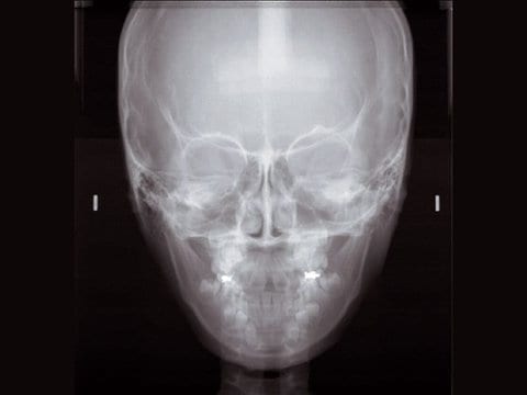
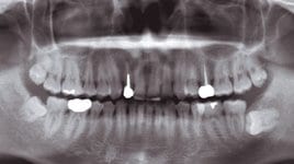
High quality images for an accurate diagnosis
High quality images for an accurate diagnosis
รายละเอียดเพิ่มเติม
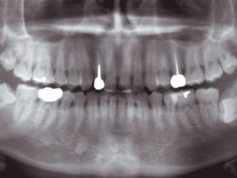
 High quality images
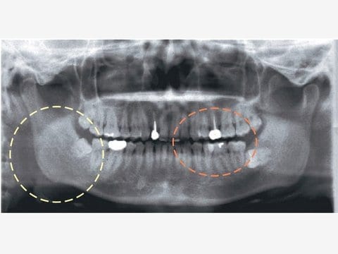
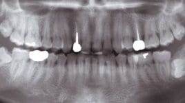
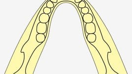
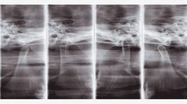
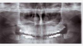
 Conventional Image
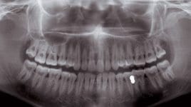
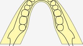
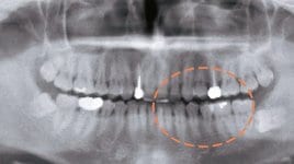
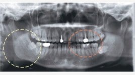
 Automatic Image Enhancer
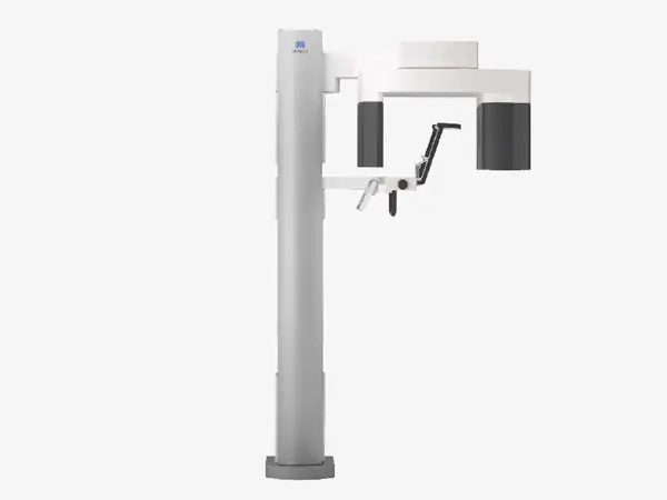
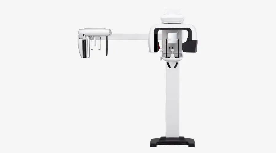
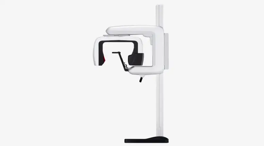
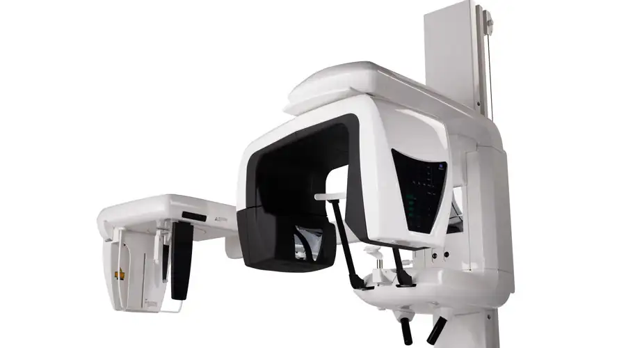
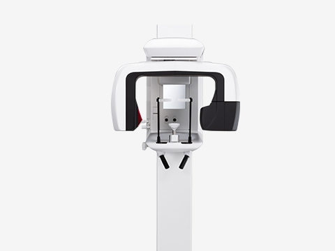
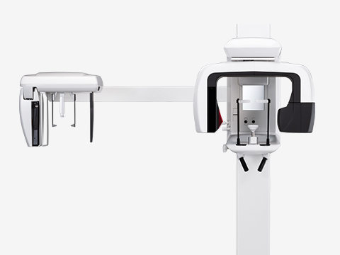
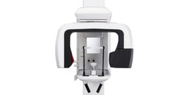
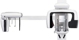
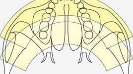
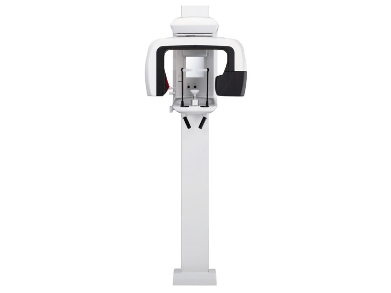
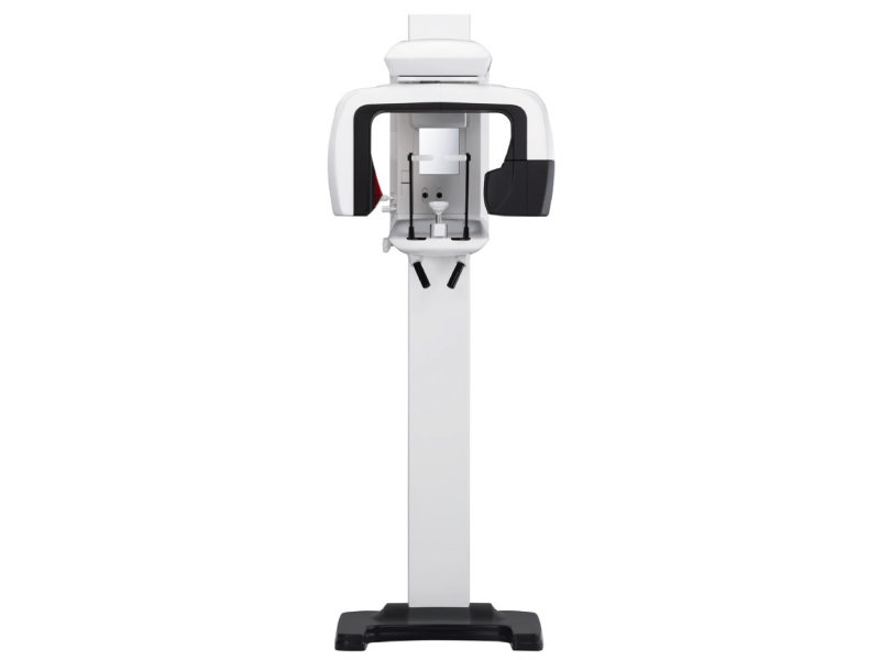
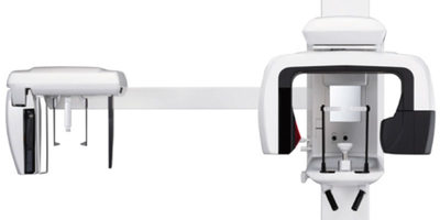
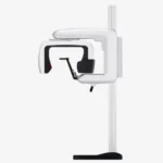
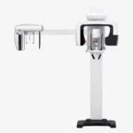
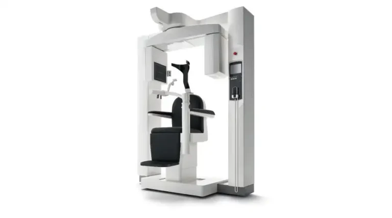
 Versatile in mode and design
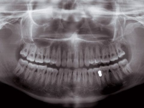
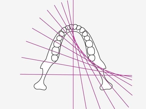
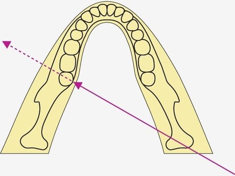
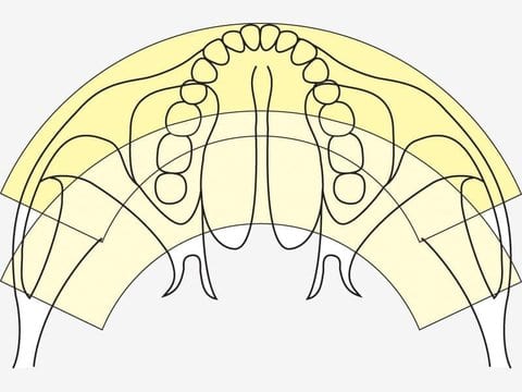
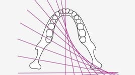
 Standard Panoramic
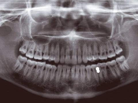
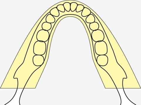
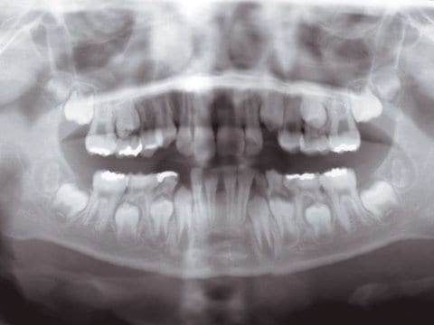
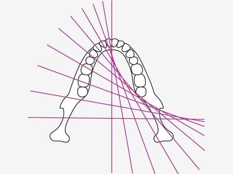
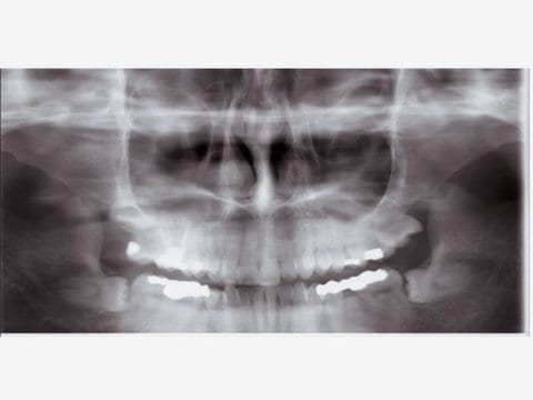
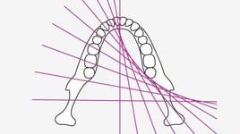
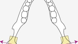
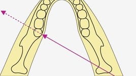
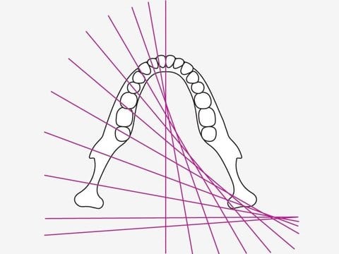 Shadow Reduction Panoramic
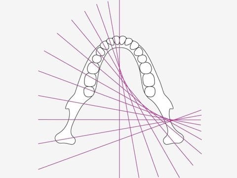
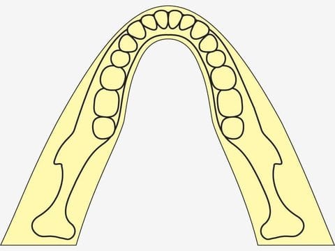
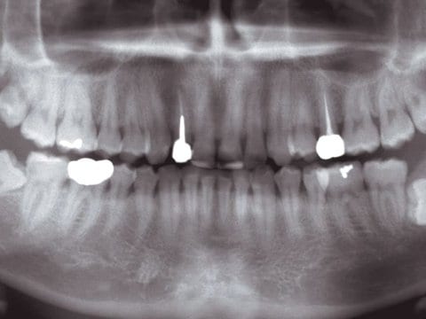
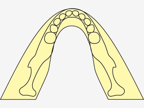
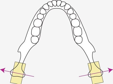
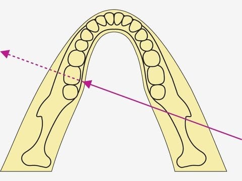
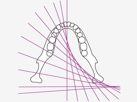
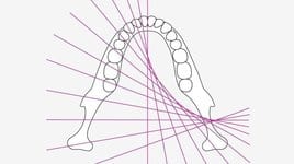
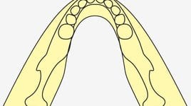
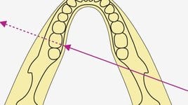
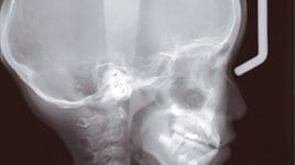
 Orthoradial Panoramic
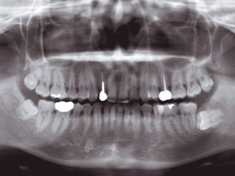
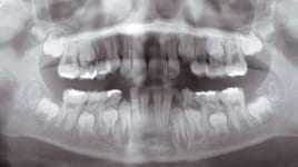
 Digital Panoramic view
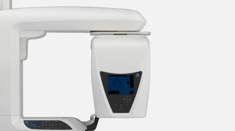
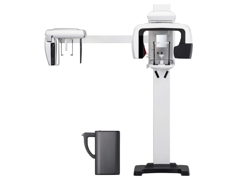
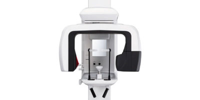
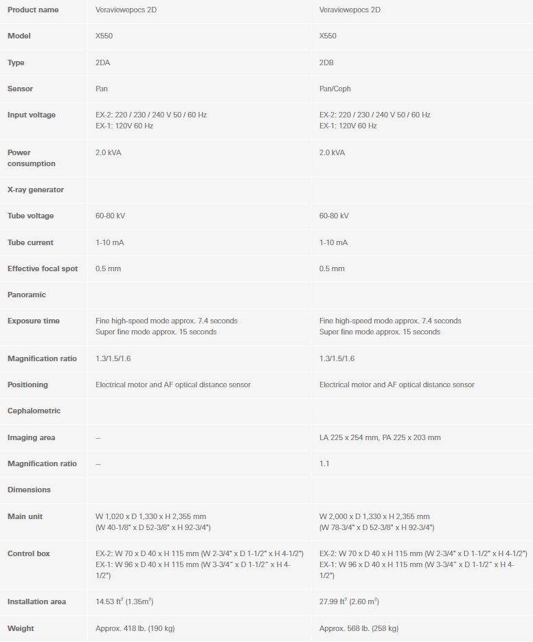
 Veraviewepocs 2D
 Veraviewepocs 2D CP
 Automatic Image Enhancer (AIE)
 Conventional Image
 Automatic Image Enhancer
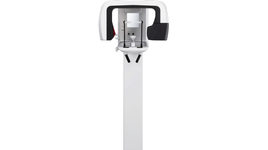 Wheelchair Support - optional wallmounted version
 Shadow Reduction Panoramic
 Orthoradial Panoramic
 Digital Panoramic - Standard Panoramic, Mag.: 1.3 x constant
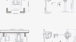
 Product sizes
 Digital Panoramic - Standard Panoramic, Mag.: 1.6 x constant
 Digital Panoramic - Pedodontic Panoramic
 Digital Panoramic - Pedodontic Panoramic
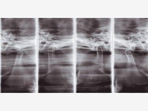
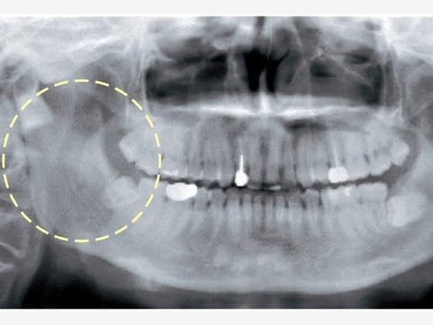
 Digital Panoramic - TMJ 4 Views, Mag.: 1.3 x constant
 Digital Panoramic - TMJ 4 Views, Mag.: 1.3 x constant
 Orthoradial Panoramic, Mag.:1.3 x constant
 Orthoradial Panoramic, Mag.:1.3 x constant
 Orthoradial Panoramic, Mag.:1.3 x constant
 Shadow Reduction Panoramic, Mag.:1.3 x constant
 Shadow Reduction Panoramic, Mag.:1.3 x constant
 Shadow Reduction Panoramic, Mag.:1.3 x constant
 Standard Panoramic, Mag.: 1.3 x constant
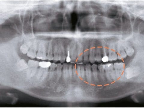
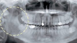
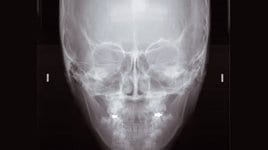
 Maxillary Sinus Panoramic, posterior Mag.: 1.5 x constant
 Maxillary Sinus Panoramic, posterior Mag.: 1.5 x constant
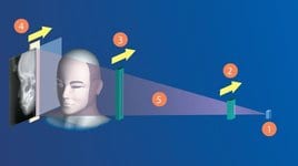 Variable image processing capabilities
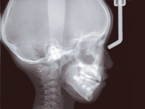
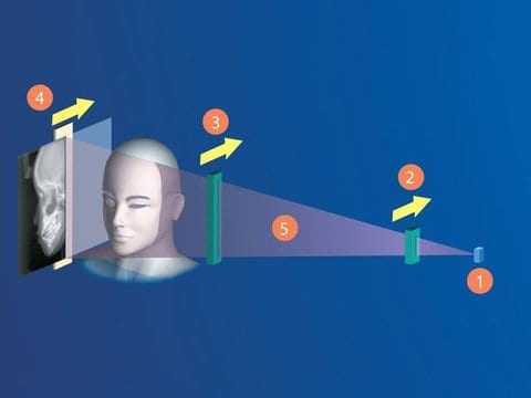
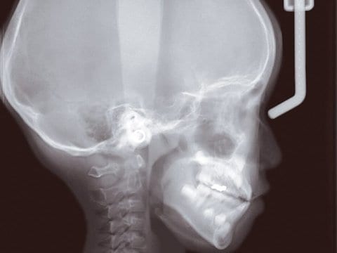
 Panoramic / Cephalometric

 EN
EN 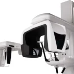
![Veraview IX R Type V-080-R EX-2 [Leather Black]](https://siamdent.kick-ring.com/wp-content/uploads/2026/05/9-1-1-768x684.webp)