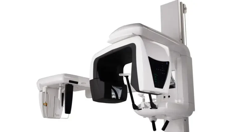
Unsurpassed image clarity
Unsurpassed image clarity
รายละเอียดเพิ่มเติม
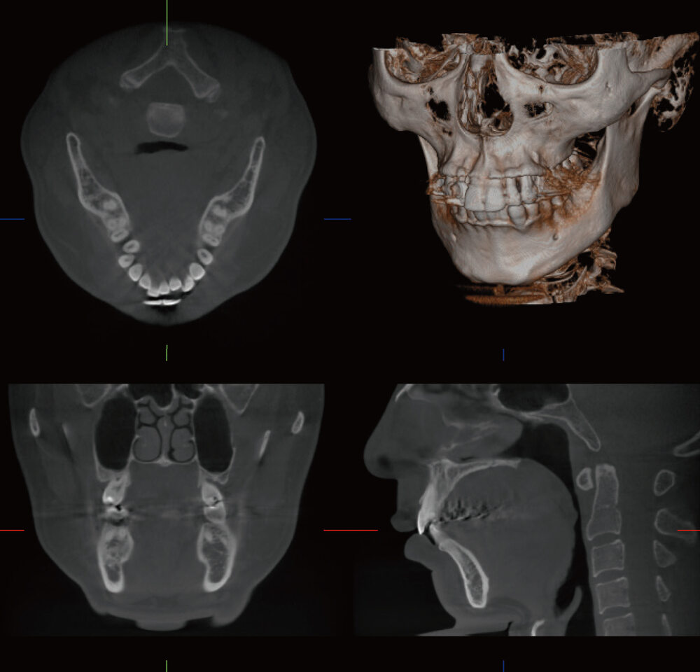
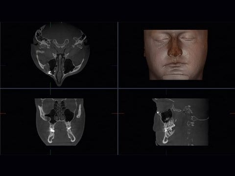
ø 170 × H 120 mm, voxel size: 250 µm
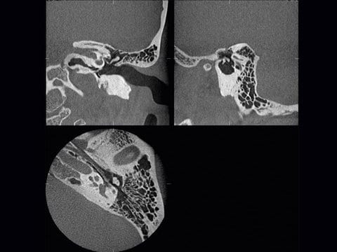
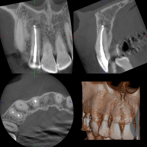 ø 40 × H 40 mm, voxel size: 80 µm
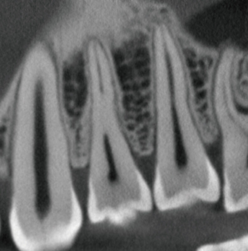 High-resolution 360°, 80μm (250μm)
 Standard-resolution 360°, 80μm
High-speed 360°, 80μm
Standard-resolution 360°, 80μm
High-speed 360°, 80μm
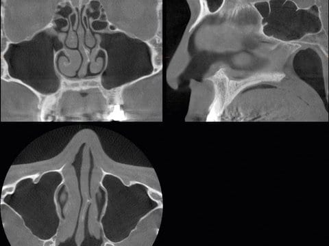
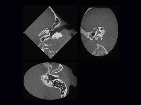
High Resolution Mode: Ø 60mm
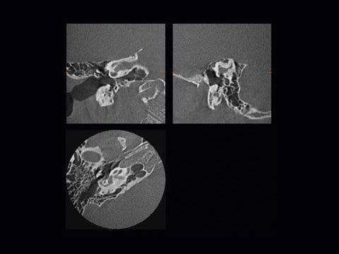
High Fidelity Mode: Ø 60 mm Zoom Reconstruction
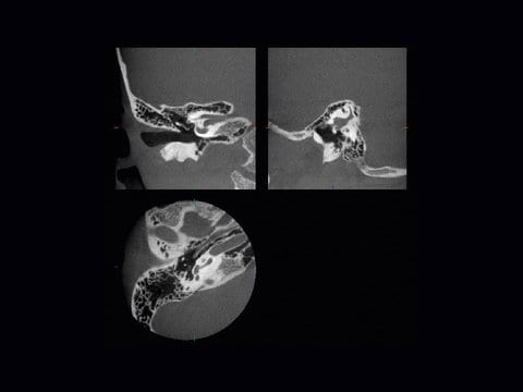
Standard mode: Ø 60 mm
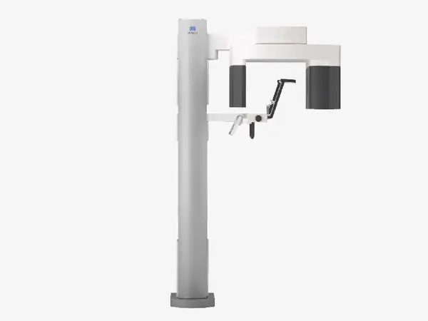
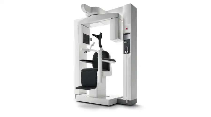
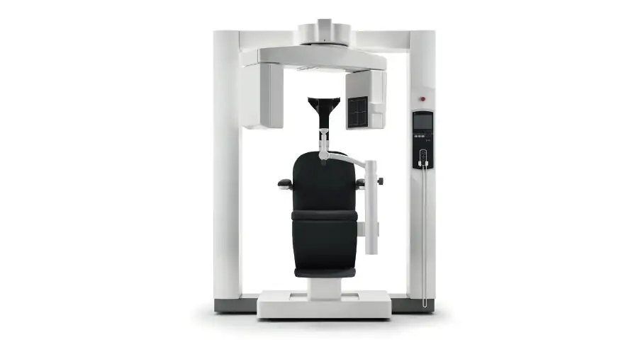
Simple, accurate positioning
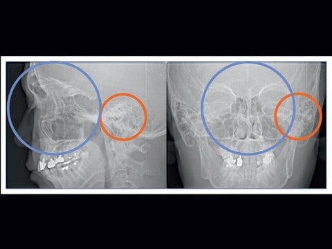
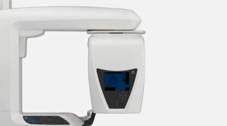
Preview Image
3D-CT image: Region of interest is well centered.
| 3D Accuitomo 170 | |
|---|---|
| Scan time in seconds | 5,9,18,30 |
| Duration of 3D exposure in seconds | 5,9,18,30 |
| Imaging in mm (WXH) | 40x40, 60x60, 80x80, 100x100, 100x50, 140x50, 140x100, 170x50, 170x120, |
| Reconstruction time | max. 5 minutes |
| Voxel size in mm³ | 0.08/0.125/0.16/0.25 |
| Lp/mm (DVT) | >2 at MTF 10% |
| Lp/mm (OPG) | N/A |
| Zoom reconstruction | yes |
| Single Images DVT | 270/530 or 900 |
| X-ray tube assembly | high frequency |
| KV/mA | 60-90 / 1-10 |
| Dosage DVT | approx. 20 µSV (4x4) |
| Dosage OPG | n.a. |
| Gray value | 14 bit |
| Image detector | Flat Panel |
| Implant planning | no software included, Dicom export for other planing programms |
| DICOM 3.0 compatible | yes |
| Storage volumina DVT | approx. 40-150 MB (to 1.4 GB rough) |
| Hardware recommendation | Network 100 Mbit/sec, Client min. Pentium 4, 1.7 GHz, 1 GB Memory |
| Included with purchase | Capture PC, Server PC, i-Dixel-software, 20" diagnostic monitor, 10x client software, One data Viewer Plus, Dicom Print, Dicom Storage |
| Name | 3D Accuitomo |
|---|---|
| Model | MCT-1 |
| Type | EX 1/2 F17 |
| Input Voltage | 100/110/120 VAC 220/230/240 VAC |
|
Power Consumption |
max. 2.0 kVA |
| Name | 3D Accuitomo |
|---|---|
| Model | MCT-1 |
| Type | EX 1/2 F17 |
| Input Voltage | 100/110/120 VAC 220/230/240 VAC |
|
Power Consumption |
max. 2.0 kVA |
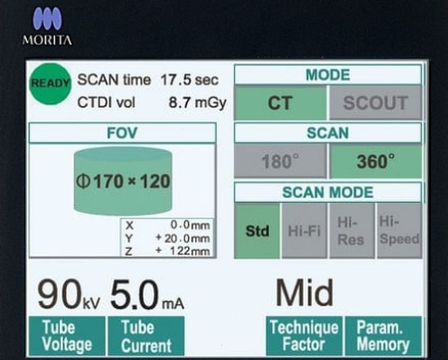
| Tube Voltage | 60-90 KV |
|---|---|
| Tube Current | 1 - 10mA (max 8 mA: Hi-Fi, Hi-Res Modes) |
| Focal Spot Size | 0.5 mm |
| Exposure Time (360°/180°) | Std-Modus: 17.5/9.0 sec. Hi-Fi-Modus: 30.8/15.8 sec. Hi-Res-Modus: 30.8/15.8 sec. Hi-Speed-Modus: 10.5/5.4 sec. |
| Field of View (Diameter × Height) | Ø 170 x120 mm Ø 170 x 50 mm Ø 140 x100 mm Ø 140 x 50 mm Ø 100 x100 mm Ø 100 x 50 mm Ø 80 x 80 mm Ø 60 x 60 mm Ø 40 x 40 mm |
| Voxel Size | 80 µm, 125 µm, 160 µm, 200 µm, 250 µm |
 Hardware Minimum Requirements for Client PC (3D X-Ray)

 EN
EN 
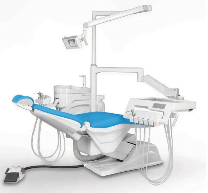
![Veraview IX R Type V-080-R EX-2 [Leather Black]](https://siamdent.kick-ring.com/wp-content/uploads/2026/05/9-1-1-768x684.webp)