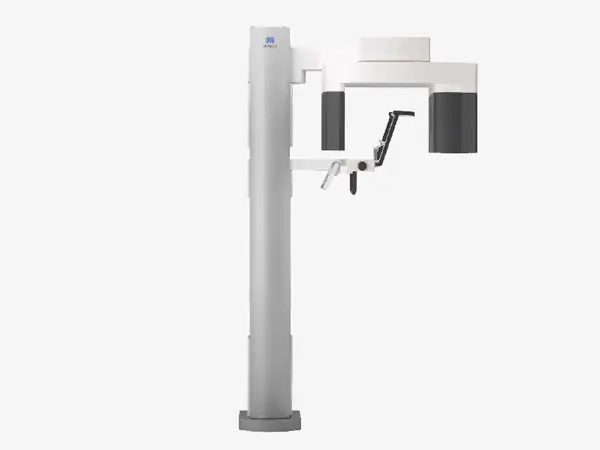
A New Frontier in X-ray Diagnostics

A New Frontier in X-ray Diagnostics
รายละเอียดเพิ่มเติม
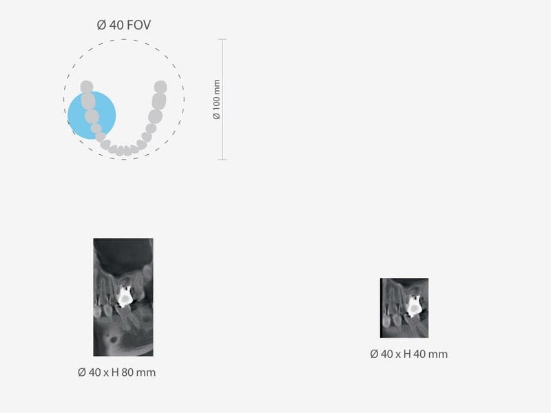
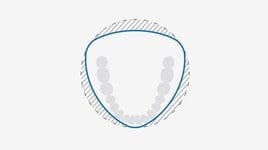
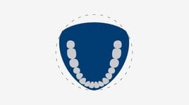
 Blue line indicates new full arch FOV, equivalent to ∅ 100 mm
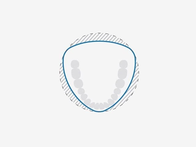 Innovative 3D Reuleaux Full Arch FOV reduces dosage.
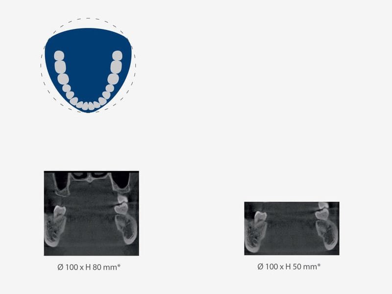
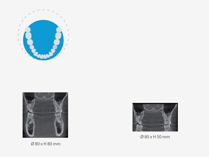
Various fields of view – Exposure Areas for Multiple Diagnostics
Innovative 3D Reuleaux Full Arch FOV reduces dosage.
Various fields of view – Exposure Areas for Multiple Diagnostics
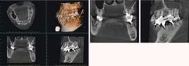
 High Resolution Images with Dose Reduction Feature
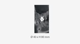
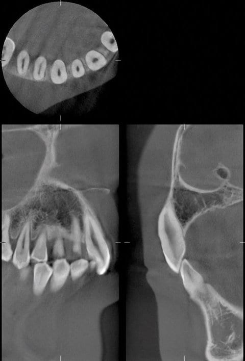 40 x 80 mm high resolution image taken in Dose Reduction Mode
 Numerous clinical cases
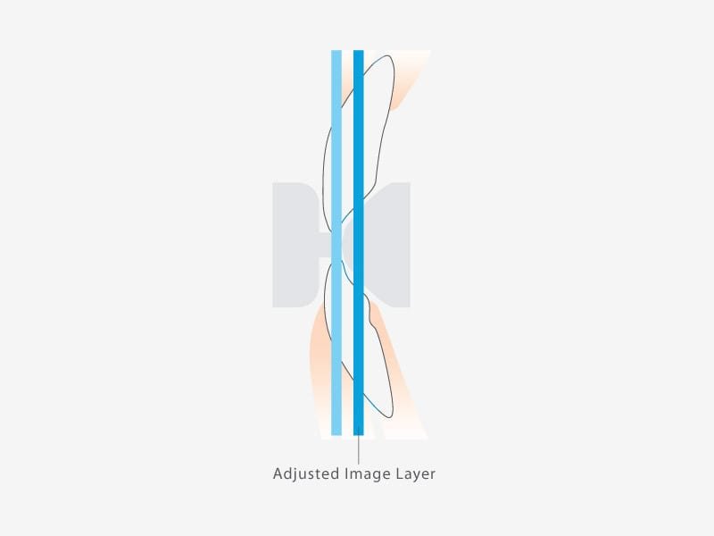
 Before Image Layer Adjustment
 Preview images are shown in the green frame to support the manipulation of image layer adjustment
 After Image Layer Adjustment
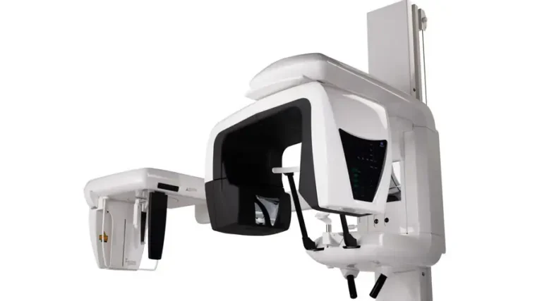
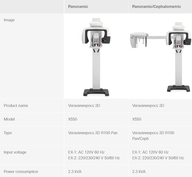
 Cephalometric Imaging
 Blue line indicates new full arch FOV, equivalent to ∅ 100 mm
 Innovative 3D Reuleaux Full Arch FOV reduces dosage
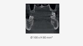
 Various Fields of View - R100 FOV
 Various Fields of View - R100 FOV
 Various Fields of View - R100 FOV
 Various Fields of View - R100 FOV
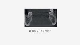
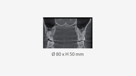
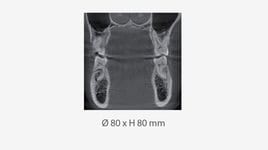 Various Fields of View - R80 FOV
 Various Fields of View - R80 FOV
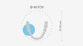 Various Fields of View - F40 FOV
 Various Fields of View - F40 FOV
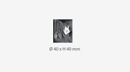 Various Fields of View - F40 FOV
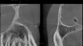 40 x 80 mm high resolution image taken in Dose Reduction Mode
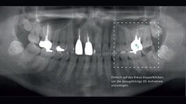 Flexibility in positioning methods - the region of interest can be positioned by the panoramic image
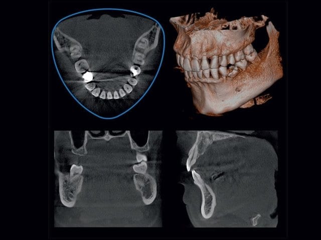
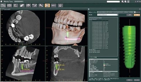
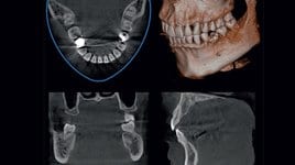
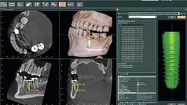 3D Images for Implant Planning with i-Dixel
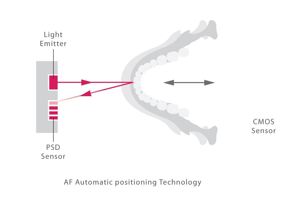
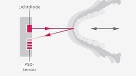 Panoramic Imaging - AF Automatic Positioning
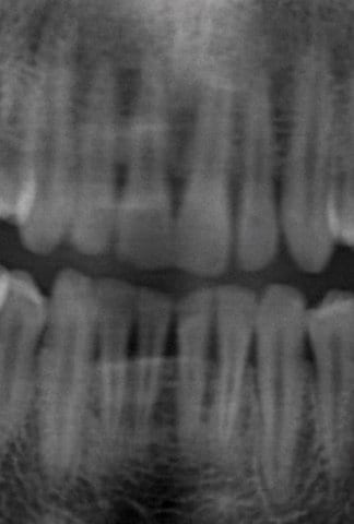
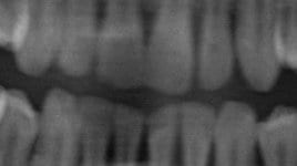 Panoramic images before Image Layer Adjustment
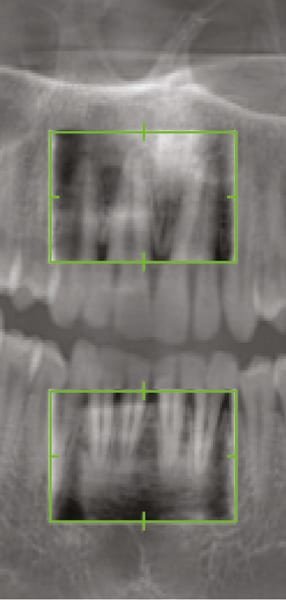
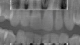 Preview images are shown in the green frame to support the manipulation of image layer adjustment
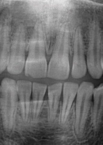
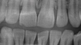 Panoramic images after Image Layer Adjustment
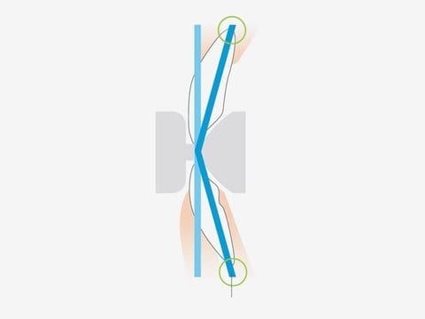
 Adjusted image layer: Two points adjustment
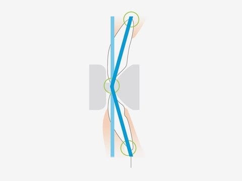
 Adjusted image layer: Three points adjustment
 Endodontics case

 EN
EN 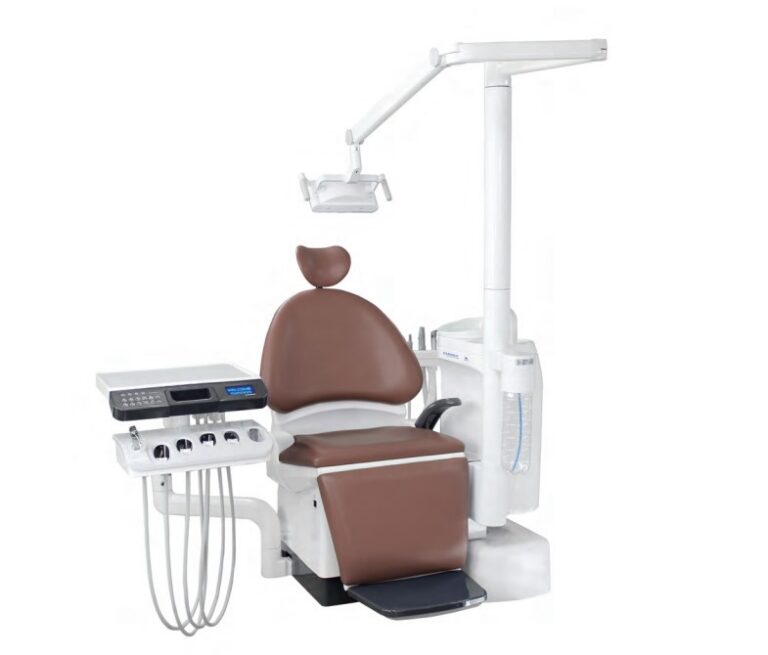
![Veraview IX R Type V-080-R EX-2 [Leather Black]](https://siamdent.kick-ring.com/wp-content/uploads/2026/05/9-1-1-768x684.webp)